Introduction
Ovarian, fallopian tube, and peritoneal (OTP) cancer is the second most common gynecological malignancy in developed countries, with an estimated prevalence in Portugal in 2020 of 9.5/100,000 women.1 Despite its relatively low incidence, OTP carcinoma is among the most lethal cancers in women, exhibiting high mortality rates.2
The most frequent histological subtype is high-grade serous carcinoma (HGSC), with approximately 75% of patients being diagnosed at advanced stages.2 Among these, 85% present with metastases confined to the peritoneal cavity.3 Breast metastases in this context are rare, estimated at 0.07%,4 and the literature addressing this clinical scenario remains limited. The main diagnostic challenge in such cases lies in distinguishing between a primary breast neoplasm and metastatic disease, to avoid unnecessary surgical intervention and to tailor treatment appropriately to the primary tumor.
There is no established standard approach for these cases. The only therapeutic option with a curative intent currently available is chemotherapy, although palliative strategies, such as radiotherapy or breast surgery, may be considered for symptomatic control. The most frequently reported approach in the literature is the combination of chemotherapy followed by local irradiation.2,3
Given the rarity and complexity of this presentation, the authors deem it relevant to report the present clinical case, despite its unfavorable outcome.
Case report
A 70-year-old caucasian female (G2P2) with a medical history of glaucoma, hypertension and depression, and no relevant family or surgical history, was referred to Gynecology for evaluation of an adnexal mass. This mass, localized in the right ovary, was heterogeneous, with 39 × 43 mm, multilocular, and had increased vascularization. Thoraco-abdominopelvic computed tomography (CT) showed no evidence of other lesions, and CA125 was 222 U/mL.
An exploratory laparotomy was performed, and intraoperative frozen section suggested HGSC of the fallopian tube. Complete surgical staging followed, including total hysterectomy, contralateral adnexectomy, infracolic omentectomy, pelvic and para-aortic lymphadenectomy, and multiple peritoneal biopsies. No macroscopic residual disease was identified. The final diagnosis was International Federation of Gynecology and Obstetrics (FIGO) stage IA HGSC of the fallopian tube. Immunohistochemistry (Fig. 1) showed positivity for paired box gene 8 (PAX 8), wingless-related integration site 1 (WT1), p16, and p53 (null-type), estrogen receptors (50-75%), and high Ki-67 (> 70%). breast cancer gene (BRCA) 1/2 testing was negative.
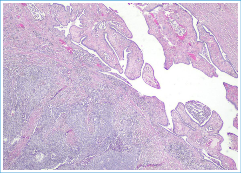
Figure 1. H&E stain (amplified ×20) section showing high-grade serous carcinoma involving the fallopian tube. Lymphovascular permeation is identifiable.
After multidisciplinary evaluation, the patient received six cycles of adjuvant carboplatin-paclitaxel chemotherapy (QT), achieving a partial response (CA125 45 U/mL; negative CT). It was decided to maintain surveillance.
Seven months later, recurrence was diagnosed: abdominal and pelvic lymphadenopathies were identified in CT (adenopathies superior to the celiac trunk and adjacent to the right iliac vessels) and raised CA125 (maximum value of 282 U/mL). The patient was asymptomatic. A biopsy confirmed inguinal metastasis. Six additional cycles of carboplatin-paclitaxel achieved partial radiological response (reduction of the volume of the adenopathies), and CA125 declined to 61.9 U/mL.
Two months later, the disease progressed (abdominopelvic lymph node regrowth). Second-line treatment was offered, which the patient refused.
Clinical surveillance was maintained and, 15 months later, bilateral cervical lymphadenopathy (in the jugulocarotid and accessory spinal chains) and CA125 elevation (332 U/mL) were observed. Cervical biopsy confirmed metastatic HGSC. Monotherapy with pegylated liposomal doxorubicin was initiated and subsequently discontinued after five cycles due to treatment-related toxicity (worsening of cardiac function), which led to clinical deterioration.
There was a subsequent elevation in CA125, and the patient reported cervical pain. An increase in the number and size of cervical lymphadenopathies was observed, with the largest nodes located on the left side, measuring up to 2 cm in maximum diameter. The case was discussed, in a multidisciplinary context, with the radiooncology team of the tertiary reference hospital: to achieve local control of the disease and symptomatic relief, it was decided to perform cervical radiotherapy (total dose of 30 Gy, in 10 fractions).
Three weeks post-radiotherapy, the patient presented at the emergency service with bowel obstruction. Clinical examination revealed, besides the cervical enlarged lymph nodes, a new breast lesion, with 4.5 cm, in the inner quadrants of the left breast, associated with skin retraction and erythema (Fig. 2). Imaging (CT) showed a suspicious retroareolar lesion (Fig. 3). Both breast and axillary biopsies confirmed metastatic HGSC, with the same immunohistochemical profile of the primary lesion, GATA3 negative, with marked lymphovascular invasion (Fig. 4).
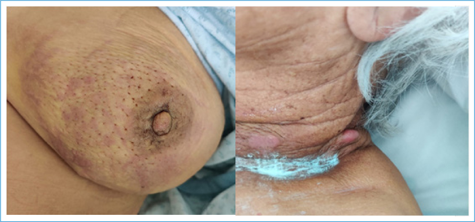
Figure 2. Left breast, portraying skin retraction and erythema, mostly on the inner quadrants; left side of the neck, showing enlarged and erythematous lymph nodes.
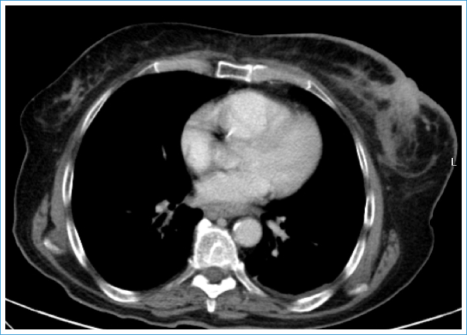
Figure 3. Thoracic computed tomography showing, on the left breast, an irregular and hyperdense retroareolar lesion, measuring approximately 6 cm, with associated skin thickening.
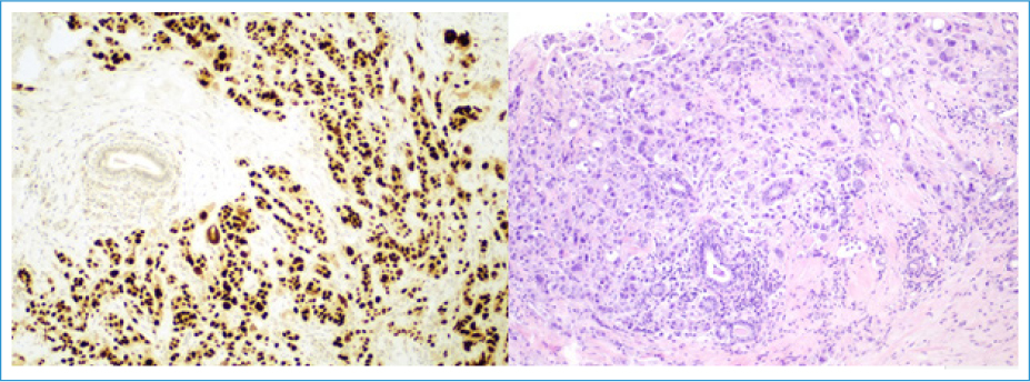
Figure 4. H&E (amplified ×100) breast lesion biopsy illustrating the high-grade serous carcinoma, with intense and diffuse expression of PAX8.
Despite supportive measures, the patient died on day 23 of hospitalization, 3½ years after initial diagnosis.
Discussion
The management of this case followed both national and international guidelines for the treatment of tubo-ovarian and peritoneal carcinoma (OTP).2,5 All decisions regarding patient monitoring and care were made within a multidisciplinary team, involving gynecology and obstetrics, medical oncology, pathology, and radiology, in collaboration with the referral radio-oncology department, ensuring appropriate surveillance.
HGSC is characterized by widespread chromosomal copy number alterations, contributing to its notable genomic instability,6 rapid proliferation, and dissemination.
Currently, no established screening strategy exists for ovarian carcinoma. In this case, diagnosis followed incidental ultrasound detection of an asymptomatic adnexal lesion. Despite early-stage disease at diagnosis and appropriate initial treatment, comprising R0 surgical resection and adjuvant chemotherapy, the patient’s disease progressed. Although post-operative CT imaging showed no evidence of residual disease, the CA125 level remained above normal (a known adverse prognostic factor). Early recurrence (within 7-8 months of chemotherapy) indicated aggressive and chemoresistant tumor behavior consistent with the literature on HGSC prognosis.
OTP carcinoma usually metastasizes through peritoneal dissemination, resulting in widespread abdominopelvic involvement. In rare cases, distant metastases may occur hematogenously or through the lymphatic system, most commonly involving the pleura, liver, lungs, and lymph nodes, particularly pelvic and para-aortic.7
Mammary involvement is extremely rare and is associated with advanced-stage disease and poor prognosis. When it occurs, HGSC is the most frequent histologic subtype implicated.7 It usually presents as a solitary mass, only 4% of cases demonstrate diffuse mammary involvement.8 This type of metastasis poses a diagnostic challenge due to its capacity to mimic a primary breast carcinoma, with non-specific or even misleading imaging findings. In our case, the lesion mimicked inflammatory breast cancer, with a rapid growth, and was identified during hospitalization for complications related to disease progression. Given the clinical context, metastatic disease was suspected, albeit rare. The role of the pathologist was critical in this scenario: immunohistochemical profiling allowed differentiation between primary breast carcinoma and metastatic OTP, revealing features consistent with the original adnexal tumor.
Progesterone and estrogen receptor expression can be present in both ovarian and breast carcinomas. However, immunohistochemical markers such as WT1, PAX8, and GATA3 allow distinguishing between primary and metastatic breast tumors: PAX8 and WT1 are expressed in approximately 88% and 72% of primary OTP carcinomas, respectively.9 In this case, the metastatic breast lesion showed positivity for WT1 and PAX8, consistent with literature findings. Conversely, GATA3, typically expressed in primary breast tumors, was negative, supporting the diagnosis of metastasis from OTP.9
Accurate lesion characterization is essential, as treatment, prognosis, and follow-up strategies differ significantly between primary breast carcinoma and metastatic OTP.
Given the patient’s extensive disease burden and deteriorating performance status, further systemic or localized breast-directed therapies were deemed inappropriate, and palliative care was prioritized.
Given its rareness, there is no standardized approach for managing breast metastases from OTP carcinoma. Fewer than 100 cases have been reported to date.3 Therapeutic strategies described include systemic chemotherapy, currently the only curative-intent option, alongside palliative modalities such as local radiotherapy, mastectomy, or lumpectomy. Surgery is typically reserved for patient’s refractory to chemotherapy, primarily for symptom relief.3 The most reported approach is the combination of chemotherapy followed by local irradiation.
Cases reported in the literature describe advanced, treatment-resistant disease, often with recurrent breast involvement after multiple chemotherapy regimens. Prognosis remains poor: reported overall survival ranges from 13 days to 3.5 years, with a 1-year survival rate of approximately 40%.10 Our patient’s survival fell within the median reported range. Contributing factors likely included advanced age,11 tumor biology, rapid progression, and poor performance status that precluded further systemic treatment.
Despite the unfavorable outcome, we present this case hoping to contribute to improving management and outcomes for similar patients. It adds to the scarce literature on breast metastasis as a late manifestation of OTP carcinoma, highlighting its diagnostic and therapeutic challenges. Further studies are needed to establish standardized diagnostic and therapeutic protocols, with effective systemic strategies, ideally based on histological and molecular profiling, to guide a multimodal treatment approach in such rare cases.
Authors’ contributions
S. Forjaz: conceptualization, writing – original draft, review and editing; L. Cunha-Silva: conceptualization, writing – original draft, review and editing; R. Coutada: clinical contribution, review; A. M. Simas: clinical contribution, review; S. Carvalho: clinical contribution, review.
Funding
None.
Conflicts of interest
None.
Ethical considerations
Protection of human subjects and animals. The authors declare that no experiments on humans or animals were performed for this research.
Confidentiality, informed consent, and ethical approval. The authors have followed their institution’s confidentiality protocols, obtained informed consent from all patients, and secured approval from the Ethics Committee. SAGER guidelines have been followed as applicable to the nature of the study.
Declaration on the use of artificial intelligence. The authors declare that no generative artificial intelligence was used in the writing or creation of the content of this manuscript.
