Introduction
Breast cancer is the most frequently diagnosed malignancy and the leading cause of cancer-related mortality among women worldwide.1 Breast carcinomas with neuroendocrine differentiation represent a rare entity, accounting for < 1% of all breast neoplasms.2,3 According to the most recent World Health Organization (WHO) classification, these neoplasms are currently included within the group of neuroendocrine neoplasms of the breast and are subdivided into well-differentiated neuroendocrine tumors, poorly differentiated neuroendocrine carcinomas, and invasive breast carcinomas of no special type with neuroendocrine differentiation.2–4
The diagnosis requires the presence of suggestive morphology in association with immunohistochemical expression of neuroendocrine markers, such as chromogranin A and synaptophysin. Exclusion of metastatic involvement of the breast from an extramammary neuroendocrine neoplasm is essential, given its distinct prognostic and therapeutic implications.4,5
Prognosis is variable and depends on the degree of differentiation, stage at diagnosis, and molecular characteristics, generally being less favorable in poorly differentiated and metastatic forms.6–9 Owing to their rarity (< 1% of breast cancers), the available literature is limited, and despite ongoing advances in research, particularly in molecular biology, specific treatment guidelines are still lacking.3 Therefore, in the absence of dedicated recommendations, therapeutic strategies are usually extrapolated from treatment algorithms for conventional invasive breast carcinoma.5,8
Case report
A 70-year-old woman with an Eastern Cooperative Oncology Group performance status of 1 was evaluated in May 2021 for a verrucous lesion located in the right inframammary fold (Fig. 1), associated with a progressively enlarging indurated nodule. The patient did not present with symptoms suggestive of carcinoid syndrome. Other than the inframammary lesion, she remained clinically asymptomatic. Breast ultrasonography performed in August 2021 revealed a mass in the lower inner quadrant of the right breast, associated with suspicious ipsilateral axillary lymphadenopathy.
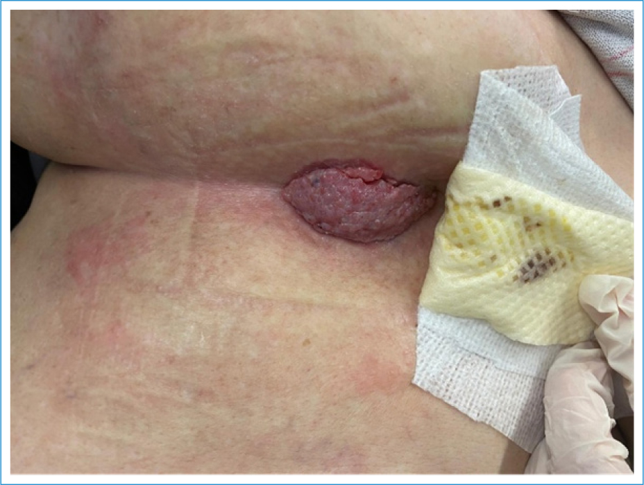
Figure 1. Verrucous lesion located in the right inframammary fold at initial presentation.
Biopsy of the cutaneous lesion demonstrated dermal infiltration by invasive breast carcinoma of no special type, with strong expression of estrogen and progesterone receptors (> 95%) and absence of human epidermal growth factor receptor-2 (HER2) expression (0). Staging computed tomography revealed bilateral pulmonary metastases and osseous involvement; these findings were confirmed by positron emission tomography (PET), which demonstrated locally advanced disease with nodal and pulmonary metastases. A lung biopsy was performed and confirmed pulmonary metastasis of carcinoma, consistent with a breast primary showing features of neuroendocrine differentiation. Immunohistochemical analysis showed diffuse positivity of the neoplastic cell population for GATA3, estrogen receptors, progesterone receptors, and synaptophysin, with absence of staining for thyroid transcription factor 1. DOTANOC PET demonstrated expression of somatostatin receptors in right axillary lymph node metastases.
In January 2022, palliative systemic treatment was initiated with letrozole 2.5 mg once daily in combination with palbociclib 125 mg once daily administered on days 1-21 of a 28-day cycle. In February 2022, long-acting release octreotide 30 mg intramuscularly every 28 days was added to the treatment regimen.
In March 2023, 13 months after treatment initiation, the patient achieved a complete clinical and radiological response, with resolution of the cutaneous lesion (Fig. 2) and no evidence of active disease on subsequent imaging. After 40 treatment cycles, she continues to tolerate therapy well, with no significant toxicities and preservation of her general condition.
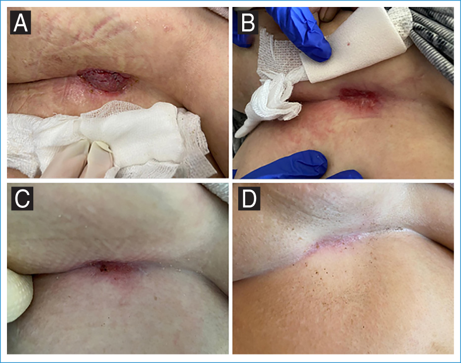
Figure 2. Evolution of the verrucous lesion during treatment. A: February 2022; B: May 2022; C: September 2022; D: March 2023.
Discussion
Breast carcinomas with neuroendocrine differentiation represent a rare entity, accounting for < 1% of breast neoplasms, and are characterized by pronounced clinical, morphological, and biological heterogeneity.2,3 The recent WHO classification update has reinforced this heterogeneity by including these neoplasms within the spectrum of neuroendocrine breast neoplasms, distinguishing well-differentiated neuroendocrine tumors, poorly differentiated neuroendocrine carcinomas, and invasive breast carcinomas of no special type with neuroendocrine differentiation.2,4 Diagnosis relies on the identification of suggestive morphology in combination with immunohistochemical expression of neuroendocrine markers, such as chromogranin A and synaptophysin, typically associated with a luminal profile, including hormone receptor (HR) expression, absence of HER2 expression, and low Ki-67 index, although other molecular subtypes have been described.4–6 The lack of uniform diagnostic criteria and morphological overlap with invasive breast carcinoma of no special type contribute to underdiagnosis and to the limited robustness of available data in the literature.4,5
From a biological standpoint, most breast carcinomas with neuroendocrine differentiation exhibit a luminal profile, with HR expression and absence of HER2 expression (HR+/HER2−), resembling classical invasive breast carcinoma.6 However, clinical and molecular studies demonstrate that this entity is not biologically homogeneous, with the degree of differentiation, proliferative index, and genetic alterations being key determinants of clinical behavior.7,9 Poorly differentiated neuroendocrine carcinomas, for instance, are characterized by frequent TP53 and RB1 alterations, higher genomic instability, and aggressive clinical behavior, and are associated with an increased risk of early metastasis and significantly poorer overall prognosis.9–11
In the absence of specific therapeutic guidelines, the management of breast carcinomas with neuroendocrine differentiation has generally been extrapolated from HR+/HER2− breast cancer treatment algorithms, particularly in the metastatic setting.8 The combination of endocrine therapy with CDK4/6 inhibitors has demonstrated consistent benefits in terms of response rate, progression-free survival, and quality of life, and currently represents the gold standard treatment for this patient subgroup.12 This approach appears particularly suitable for tumors with a luminal profile and less aggressive biological behavior, such as well-differentiated neuroendocrine tumors and invasive carcinomas with neuroendocrine differentiation.
A distinctive and clinically relevant feature of these tumors is the expression of somatostatin receptors, reported in approximately 50-70% of cases.5,13 Functional identification of these receptors through molecular imaging techniques, such as DOTANOC PET, not only allows for improved disease characterization but also enables consideration of additional targeted therapeutic strategies. Somatostatin analogs, namely octreotide and lanreotide, have demonstrated antiproliferative, antisecretory, and disease-stabilizing effects in well-differentiated neuroendocrine neoplasms, and are associated with prolonged disease control and good treatment tolerability.13,14 Although evidence in breast carcinoma with neuroendocrine differentiation is limited and primarily based on small case series and reports, the physiological rationale for their use appears sound.13
In the present case, histological confirmation of neuroendocrine differentiation, combined with functional demonstration of somatostatin receptor expression, enabled a truly personalized therapeutic approach. The combination of palbociclib and letrozole, in line with recommendations for metastatic HR+/HER2− breast carcinoma, together with octreotide therapy, resulted in a complete and sustained clinical and radiological response over 40 treatment cycles, with excellent tolerability and preservation of the patient’s functional status. This outcome suggests a potential synergistic effect between endocrine therapy, cell cycle inhibition, and modulation of neuroendocrine signaling, translating into prolonged metabolic and tumor control.
Notably, in this patient, neuroendocrine differentiation was identified on histological evaluation of a metastatic lesion, underscoring the importance of comprehensive pathological reassessment not only of the primary breast tumor but also of metastatic sites whenever feasible. This finding highlights the potential biological heterogeneity between primary and metastatic disease and emphasizes that biopsy of metastatic lesions may provide additional diagnostic and predictive information with direct implications for therapeutic decision-making and treatment personalization.
Overall, this case underscores the importance of comprehensive histological, molecular, and functional characterization in breast carcinoma with neuroendocrine differentiation, enabling the identification of patient subgroups that may benefit from integrated and targeted therapeutic strategies. Despite the inherent limitations of a single case report, the sustained response observed supports the need for prospective studies and multicenter registries to clarify the role of somatostatin analogs in this context and to define more specific therapeutic approaches for this rare entity.
Conclusion
Breast carcinoma with neuroendocrine differentiation is rare and exhibits a variable prognosis. Histological and functional characterization using DOTANOC PET can identify important therapeutic targets. The combination of somatostatin analogs with endocrine therapy and CDK4/6 inhibitors demonstrated a prolonged and clinically meaningful response, illustrating the potential of integrated, targeted, and personalized approaches in breast cancer.
Funding
None.
Conflicts of interest
None.
Ethical considerations
Protection of human subjects and animals. The authors declare that no experiments on humans or animals were performed for this research.
Confidentiality, informed consent, and ethical approval. The authors have followed their institution’s confidentiality protocols, obtained informed consent from all patients, and secured approval from the Ethics Committee. SAGER guidelines have been followed as applicable to the nature of the study.
Declaration on the use of artificial intelligence. The authors declare that no generative artificial intelligence was used in the writing or creation of the content of this manuscript.
