
A 71-year-old male was referred to our hospital for hepatology assessment due to suspected chronic liver disease. His past medical history was unremarkable, except for moderate chronic alcohol consumption. Physical examination and vital signs were within normal limits. Laboratory evaluation revealed normal alpha-fetoprotein levels, normal liver function tests, and normal prothrombin time.
Considering the patient’s medical history and the unfavorable body habitus for abdominal ultrasound evaluation, contrast-enhanced computed tomography (CECT) of the abdomen was performed. A lesion was identified in segment VII of the liver, demonstrating arterial phase enhancement (Fig. 1), with washout in the portal venous phase (Fig. 2). The right adrenal gland was not clearly visualized (Fig. 2). No intralesional macroscopic fat or imaging signs of chronic liver disease were detected on CECT. To better characterize the hepatic lesion, magnetic resonance imaging (MRI) with extracellular gadolinium-based contrast was requested, showing signal intensity loss on in-phase and out-of-phase T1-weighted sequences (Figs. 3 and 4), consistent with the presence of intralesional microscopic fat.
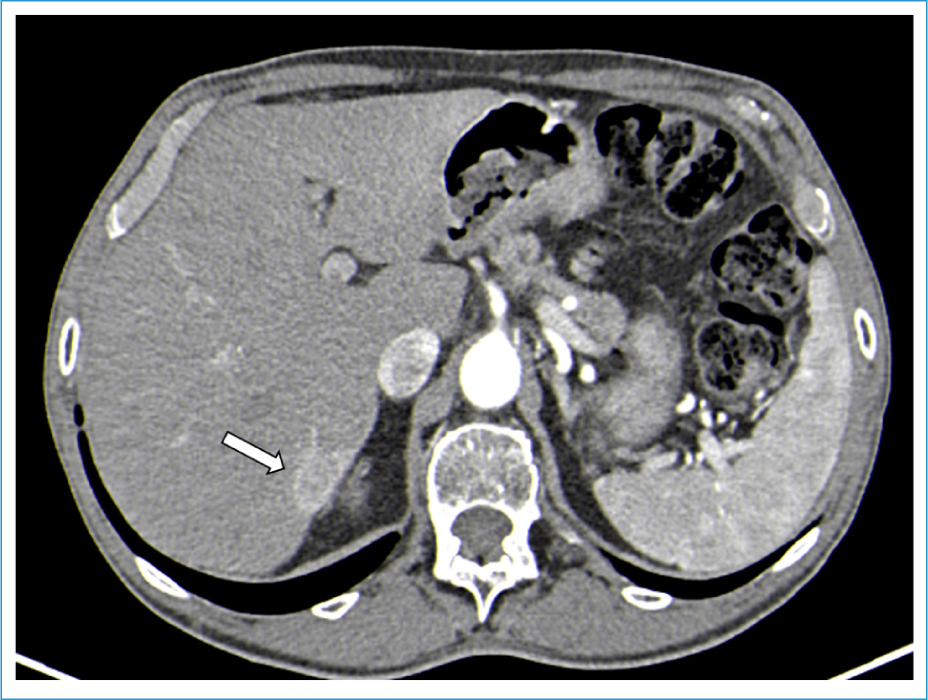
Figure 1. Computed tomography scan, axial arterial phase. The white arrow shows a hypervascular nodule in the VII segment measuring 19 mm.
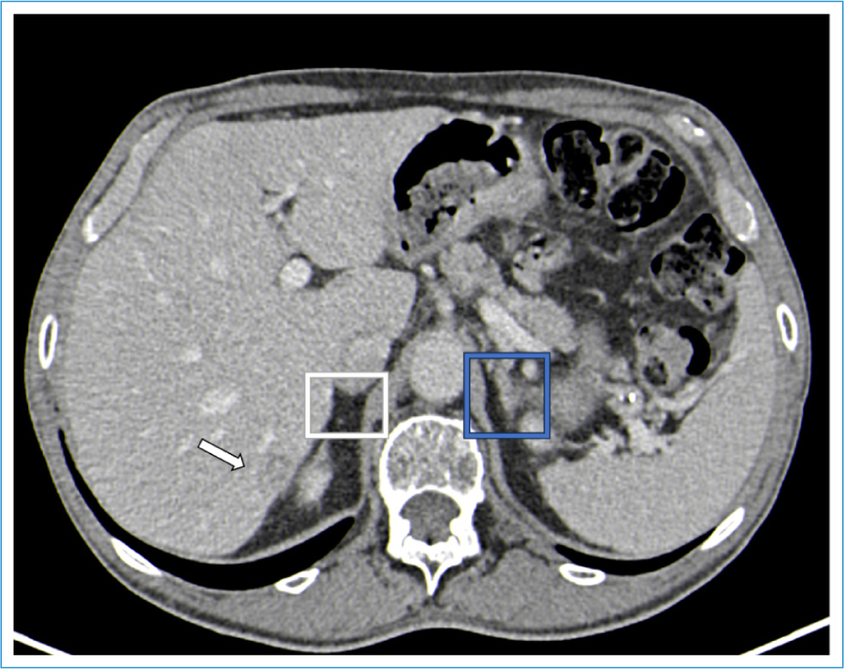
Figure 2. Computed tomography scan, axial portal phase. The white arrow shows washout; white square shows where the right adrenal gland was supposed to be observed; the blue square shows the left adrenal gland.
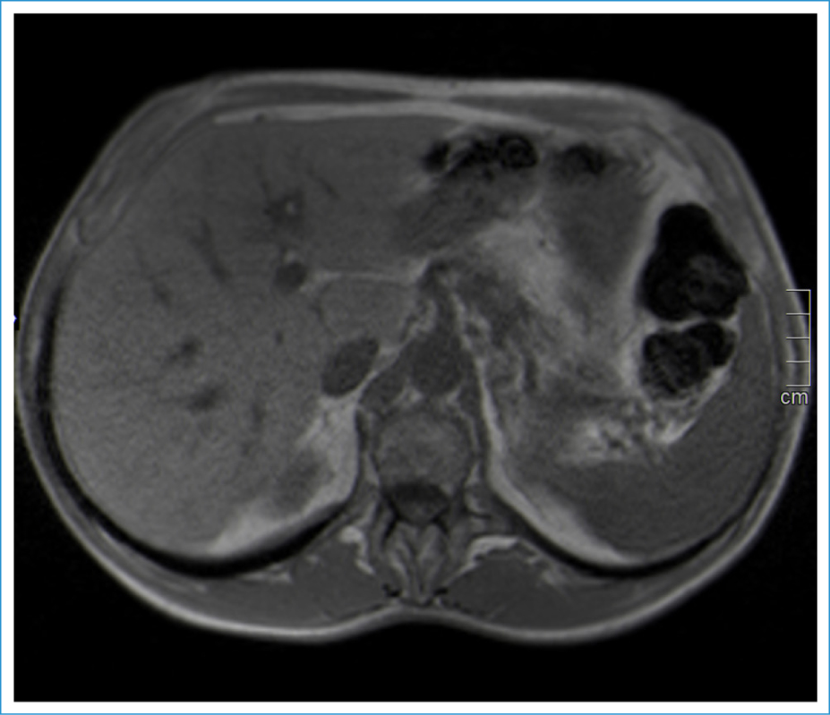
Figure 3. Magnetic resonance T1 in-phase sequence axial.
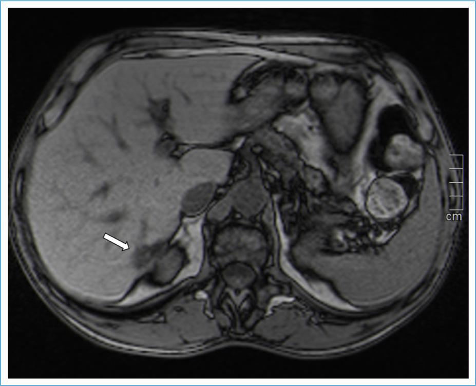
Figure 4. Magnetic resonance T1 out-of-phase sequence axial. The white arrow shows signal drop of the subcapsular lesion in segment VII.
Hepatocellular carcinoma (HCC) was initially suspected; however, after multidisciplinary discussion, follow-up imaging at 3 months was recommended. This decision was based on the unclear visualization of the right adrenal gland, raising the possibility of a hepatic adrenal rest tumor (HART). Subsequent imaging demonstrated lesion stability, supporting the diagnosis of HART.
Adrenal rest tumors are defined as collections of aberrant adrenocortical tissue located outside the adrenal gland.1,2 They are rare entities, most commonly found in the abdomen or pelvis, with the kidneys being the most frequent location. HARTs are extremely rare.1,2
Some lesions may be hormonally active and present with endocrine syndromes,1 and immunohistochemical studies can confirm steroidogenic capability.1 Malignant transformation is exceedingly rare.2
On imaging, HARTs usually appear as well-defined, round subcapsular masses in the posterior right lobe of the liver.3 They typically present as hypervascular, fat-containing lesions.3 Intralesional fat may be detected on CECT or ultrasound, but it is more accurately characterized by MRI.3 The differential diagnosis includes fat-containing HCC, focal nodular hyperplasia, hepatic adenoma, hepatic angiomyolipoma, hepatic lipoma, and primary hepatic or metastatic liposarcoma.3 Another important imaging feature is hypervascularity with early arterial enhancement and washout, which makes differentiation from HCC particularly challenging, especially in cirrhotic patients.2,4
Histologically, HARTs resemble adrenal cortical tumors and are composed of low columnar or cuboidal clear cells, typically rich in lipid content and arranged in cord-like patterns separated by thin fibrous septa.1 Immunohistochemistry usually demonstrates positivity for Melan-A, confirming adrenal origin, and negativity for HepPar-1 and PAX8, excluding hepatocellular and renal origin, respectively.5
Although no pathognomonic imaging findings exist, HART should be considered in the differential diagnosis of hypervascular, fat-containing hepatic lesions located in segment VII, particularly in patients with normal alpha-fetoprotein levels and an unidentifiable right adrenal gland. Recognizing this entity is essential to avoid unnecessary hepatectomy or liver transplantation without curative intent, given the benign nature of HART.
Authors’ contributions
All the authors mentioned above contributed to the preparation of this article, namely in the case follow-up, image collection, literature review, and manuscript writing.
Funding
None.
Conflicts of interest
None.
Ethical considerations
Protection of humans and animals. The authors declare that no experiments involving humans or animals were conducted for this research.
Confidentiality, informed consent, and ethical approval. The authors have followed their institution’s confidentiality protocols, obtained informed consent from patients, and received approval from the Ethics Committee. The SAGER guidelines were followed according to the nature of the study.
Declaration on the use of artificial intelligence. The authors declare that no generative artificial intelligence was used in the writing of this manuscript.
