Pulmonary pseudotumors are relatively common radiologic findings. Most frequently caused by a loculated pleural effusion confined to the pleural fissures, other etiologies have been described, such as inflammation, atelectasis, fat, mucoid, or calcifying impaction, among others.1
We report the case of a 62-year-old man, previously a house-constructor, current smoker (45 pack-years), with a known history of hypertension and alcohol-related liver cirrhosis. He was referred to our department with a diagnosis of bilateral pneumonia with no signs of heart failure or ascites.
Chest X-ray (Fig. 1) showed a suspicious right paratracheal fusiform opacity and features suggestive of pulmonary congestion. Computed tomography of the chest (Fig. 2) revealed the presence of an accessory azygos lobe (a rare anatomical variation) with a loculated pleural effusion in the respective fissure (forming an “hourglass” shape).
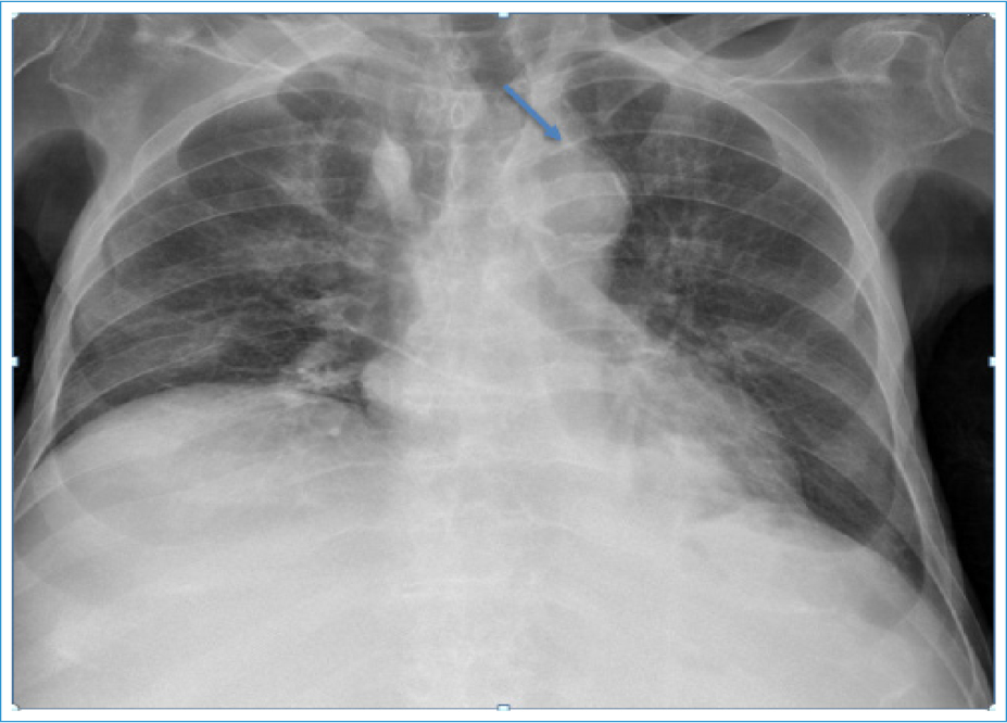
Figure 1. Chest X-ray demonstrating a suspicious right paratracheal fusiform opacity (arrow).
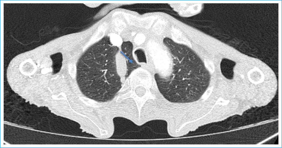
Figure 2. Computed tomography of the chest showing an accessory lobe of the azygos vein (anatomical variation) with a loculated pleural effusion within the azygos fissure (arrow).
The image had significant variation after diuretic treatment, and a completed resolution was registered. This case aligns with the classic concept of “pulmonary pseudotumor”, sometimes also described as “phantom tumor”.1
These types of images are relatively common in the pulmonary major fissures,2–5 and they may raise diagnostic difficulties, particularly among less trained clinicians.1,5 In our bibliographic research, we could not find previous case-reports of azygos fissure pseudotumor, hence the relevance of the image reported above.
Authors’ contributions
C. Honrado-Martins: conception, drafting, and critical review; G. Castro-Gomes: critical review and approval of the final version.
Funding
None.
Conflicts of interest
None.
Ethical considerations
Protection of human subjects and animals. The authors declare that no experiments on humans or animals were performed for this research.
Confidentiality, informed consent, and ethical approval. The authors have followed their institution’s confidentiality protocols, obtained informed consent from all patients, and secured approval from the Ethics Committee. SAGER guidelines have been followed as applicable to the nature of the study.
Declaration on the use of artificial intelligence. The authors declare that no generative artificial intelligence was used in the writing or creation of the content of this manuscript.
